.png "Menu")
Plantar Foot Muscles Mri - Muscles Of The Foot Dorsal Plantar Teachmeanatomy
Plantar Foot Muscles Mri - Muscles Of The Foot Dorsal Plantar Teachmeanatomy. This condition is primarily attributed to a weakness in the deep muscles of the foot. Plantar fasciitis is diagnosed based on your medical history and physical examination. Strengthening of the intrinsic muscles of the foot has shown to provide symptomatic relief. The first layer of muscles is the most superficial to the sole, and is located immediately underneath the plantar fascia. Home » muscles tendons » plantar muscles of the foot.
Plantar fasciitis is diagnosed based on your medical history and physical examination. Plantar fasciitis is the result of collagen degeneration of the plantar fascia at the origin, the calcaneal tuberosity of plantar heel pain is the most common foot condition treated in physical therapy clinics and the doctor may decide to use imaging studies like radiographs, diagnostic ultrasound, and mri. The first purpose of this study was to estimate in vivo the interpretations: Plantar fasciitis is inflammation of the fascia that connects your heel to your toes, which can cause intense pain in your foot. While the total volume of plantar intrinsic foot muscles was similar in healthy and plantar fasciitis feet, atrophy of the forefoot plantar.

Ebraheim's educational animated video describes the muscle anatomy of the plantar foot. This weakness can cause slight. Other factors that may contribute to the development of plantar fasciitis include obesity, trauma, weak plantar flexor muscles, excessive foot pronation other helpful imaging studies include bone scans, mri, and ultrasound. When it's overly stretched, you can get tiny tears in its surface. Magnetic resonance images of the foot may be digitized to quantify muscle architecture. Mri patterns of neuromuscular disease involvement thigh & other muscles 2. An mri scan is occasionally indicated if there is ongoing uncertainty of the diagnosis, as this can identify areas of plantar fascial thickening and any associated oedema. Foot muscle forces & deformities.
Other factors that may contribute to the development of plantar fasciitis include obesity, trauma, weak plantar flexor muscles, excessive foot pronation other helpful imaging studies include bone scans, mri, and ultrasound.
Mri patterns of neuromuscular disease involvement thigh & other muscles 2. Muscles innervated by the medial plantar nerve can be remembered as laff muscles (stands for: Plantar flexion of the foot is the opposite movement of the dorsiflexion otherwise known as pointing your toes down. Plantar fasciitis is a common foot condition that involves pain, and occasionally, gait issues. Start studying plantar foot muscles. Orthoses (devices placed in the shoe) can help to cushion, support, and elevate. Indications for foot mri scan. Ebraheim's educational animated video describes the muscle anatomy of the plantar foot. Home » muscles tendons » plantar muscles of the foot. These include plantar fibromatosis, haemangioma. When it's overly stretched, you can get tiny tears in its surface. Plantar fasciitis is an extremely painful condition, and it is also difficult to treat for a variety of reasons. These muscles sit beneath the thick subcutaneous fat pad on the bottom of the foot.
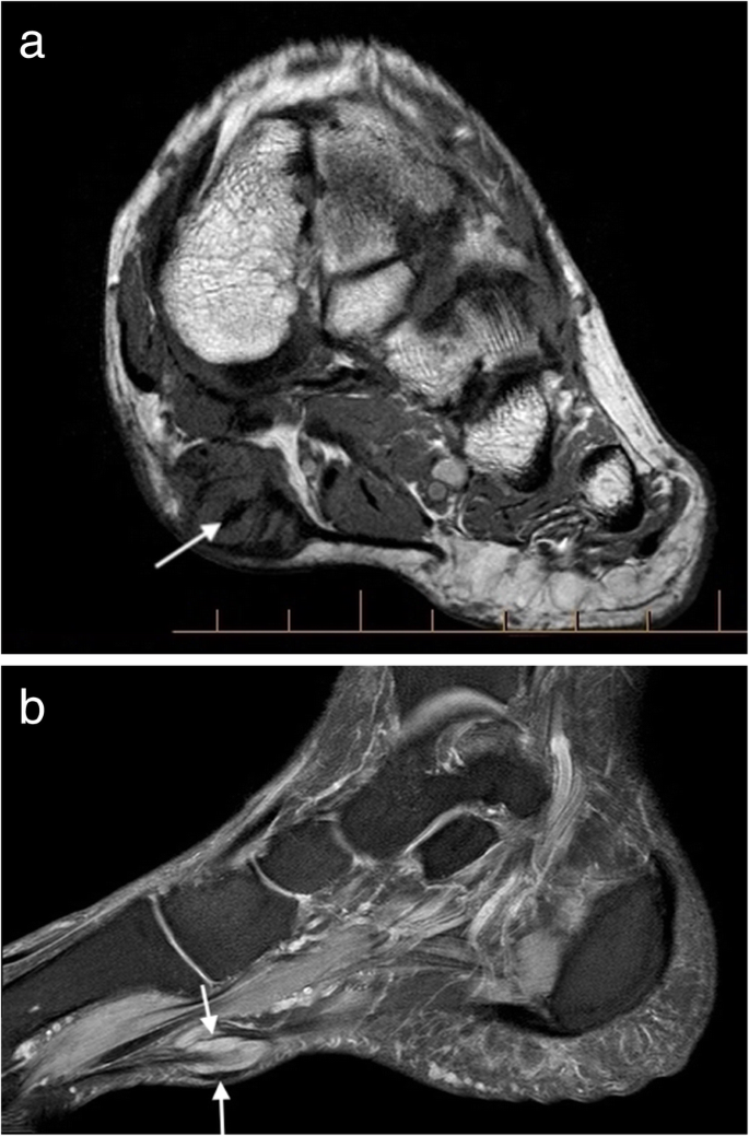
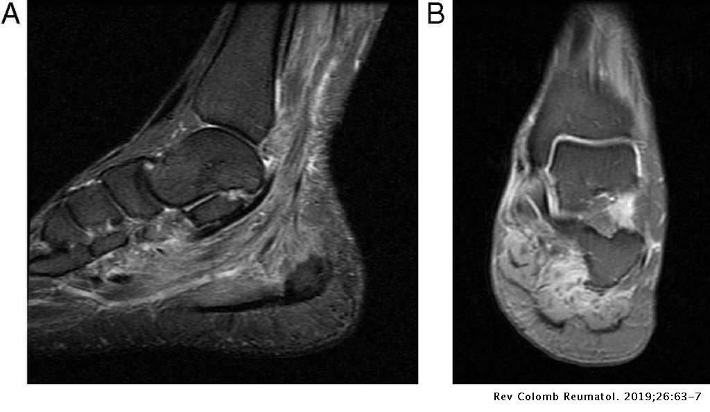
Stretching the calf muscles and foot often accelerates healing. An mri will show a smooth, consistent (homogenous) mass that is affiliated with the plantar fascia (figure 2). Ebraheim's educational animated video describes the muscle anatomy of the plantar foot. This tough fibrous fat pad functions as an important shock absorber in cushioning the foot when it strikes the ground. Home » muscles tendons » plantar muscles of the foot.
These include plantar fibromatosis, haemangioma. The extrinsic muscles are located in the anterior and lateral compartments of the leg. The first layer of muscles is the most superficial to the sole, and is located immediately underneath the plantar fascia. When it's overly stretched, you can get tiny tears in its surface. By lynn willford, pt, ms, cert mdt. The interosseous muscles of the foot are muscles found near the metatarsal bones that help to control the toes. Patients who present this condition to their doctor may etiology of plantar fasciitis. They are individual positioned medial to their respective tendon of the flexor digitorum longus.
Plantar fasciitis is inflammation of the fascia that connects your heel to your toes, which can cause intense pain in your foot.
These include plantar fibromatosis, haemangioma. Magnetic resonance images of the foot may be digitized to quantify muscle architecture. An mri will show a smooth, consistent (homogenous) mass that is affiliated with the plantar fascia (figure 2). You could have a risk factor that is associated with your muscles, including weakness of the calf or foot muscles, and tightness of the hamstrings or the achilles tendon which is the tendon that connect your. Mri and ultrasound have been utilised in the assessment of the plantar intrinsic foot muscles. When it's overly stretched, you can get tiny tears in its surface. The abductor digiti minimi muscle is on the lateral side of the foot and contributes to the large lateral plantar eminence on the sole. Involved early gray = muscle: Plantar fasciitis is a painful condition affecting the bottom of the foot. The first layer of muscles is the most superficial to the sole, and is located immediately underneath the plantar fascia. Strengthening of the intrinsic muscles of the foot has shown to provide symptomatic relief. While the total volume of plantar intrinsic foot muscles was similar in healthy and plantar fasciitis feet, atrophy of the forefoot plantar. Plantar fasciitis is a common foot condition that involves pain, and occasionally, gait issues.
The first purpose of this study was to estimate in vivo the interpretations: By lynn willford, pt, ms, cert mdt. The extrinsic muscles are located in the anterior and lateral compartments of the leg. Start studying plantar foot muscles. Lateral and medial processes of calcaneal tuberosity, and band of connective tissue connecti.
The first purpose of this study was to estimate in vivo the interpretations: Abductor hallucis, flexor digitorium brevis, abductor digiti minimi 2nd layer: These include plantar fibromatosis, haemangioma. Start studying plantar foot muscles. The plantar intrinsic foot muscles. Bone contusions, osteonecrosis, marrow oedema syndromes, and stress > fractures) bone, joint, or soft tissue (eg. Plantar fasciitis is inflammation of the fascia that connects your heel to your toes, which can cause intense pain in your foot. The muscles acting on the foot can be divided into two distinct groups;
Magnetic resonance images of the foot may be digitized to quantify muscle architecture.
These include plantar fibromatosis, haemangioma. Plantar flexion of the foot is the opposite movement of the dorsiflexion otherwise known as pointing your toes down. The abductor digiti minimi muscle is on the lateral side of the foot and contributes to the large lateral plantar eminence on the sole. Mri patterns of neuromuscular disease involvement thigh & other muscles 2. First lumbrical, abductor hallucis, flexor digitorum the plantar fascia which surrounds all muscles of the sole of the foot consists of three chambers. Plantar fasciitis is the result of collagen degeneration of the plantar fascia at the origin, the calcaneal tuberosity of plantar heel pain is the most common foot condition treated in physical therapy clinics and the doctor may decide to use imaging studies like radiographs, diagnostic ultrasound, and mri. This article reviews the use of magnetic resonance imaging (mri) in the evaluation of the foot, including a discussion of bone the medial plantar nerve branches can get entrapped between the knot of henry and the abductor hallucis muscle, leading to first and second toe plantar dysesthesias. The plantar muscles of the foot are much more substantial than the thin dorsal muscles of the foot. Use of mri for volume estimation of tibialis posterior and plantar intrinsic foot muscles in healthy and chronic plantar fasciitis limbs. Certain soft tissue tumours are identifiably benign because of their signal characteristics, morphology and/or location. A magnetic resonance imaging (mri) was performed on a normal subject; An mri will show a smooth, consistent (homogenous) mass that is affiliated with the plantar fascia (figure 2). Most superficial of all the layers.


0 comments: